Writer and PhD candidate Taylor Mabe works with his faculty mentor Dr. Jianjun Wei in the UNCG Department of Nanoscience, part of the Joint School of Nanoscience and Nanoengineering. His poster, “Development and fabrication of a handheld point-of-care sensor for disease diagnosis,” took 1st place in Natural, Physical and Mathematical Sciences at the 2016 Graduate Research and Creativity Expo.
Imagine the benefits to mankind if we could do a chemical analysis anywhere in the world at any time. Examining a soldier’s blood for toxins on the battlefield, emotional STD screening in the privacy of the home, or bedside analysis for patients in remote regions – all become possibilities.
My research, guided by Dr. Jianjun Wei, focuses on fabricating devices that can turn such possibilities into a reality. These miniaturized devices, aptly called biosensors, analyze biomarkers, which are molecules made in response to changes in the body, such as a disease.
One issue with disease diagnostics is that in order to identify a disease early on, prior to the onset of symptoms, we need to detect biomarkers when they are at low concentrations. This is truly finding a needle in a haystack. A single droplet of blood contains 5 million red blood cells and an array of proteins. Out of that complexity we are looking for a single type of protein. It’s like fishing the Atlantic Ocean for a single, specific fish.
Complex lab samples, like blood, have a lot of “stuff” in them and need to be separated, so each component can be detected. This is currently done using high-pressure liquid chromatography mass spectrometry. The bulky instruments are not portable, destroy the sample, and require a lot of energy and expertise to operate.
The devices we are designing can conduct the same analyses, but at a square inch in size our instruments offer portability, low cost, faster assay times, and a smaller sample requirement.
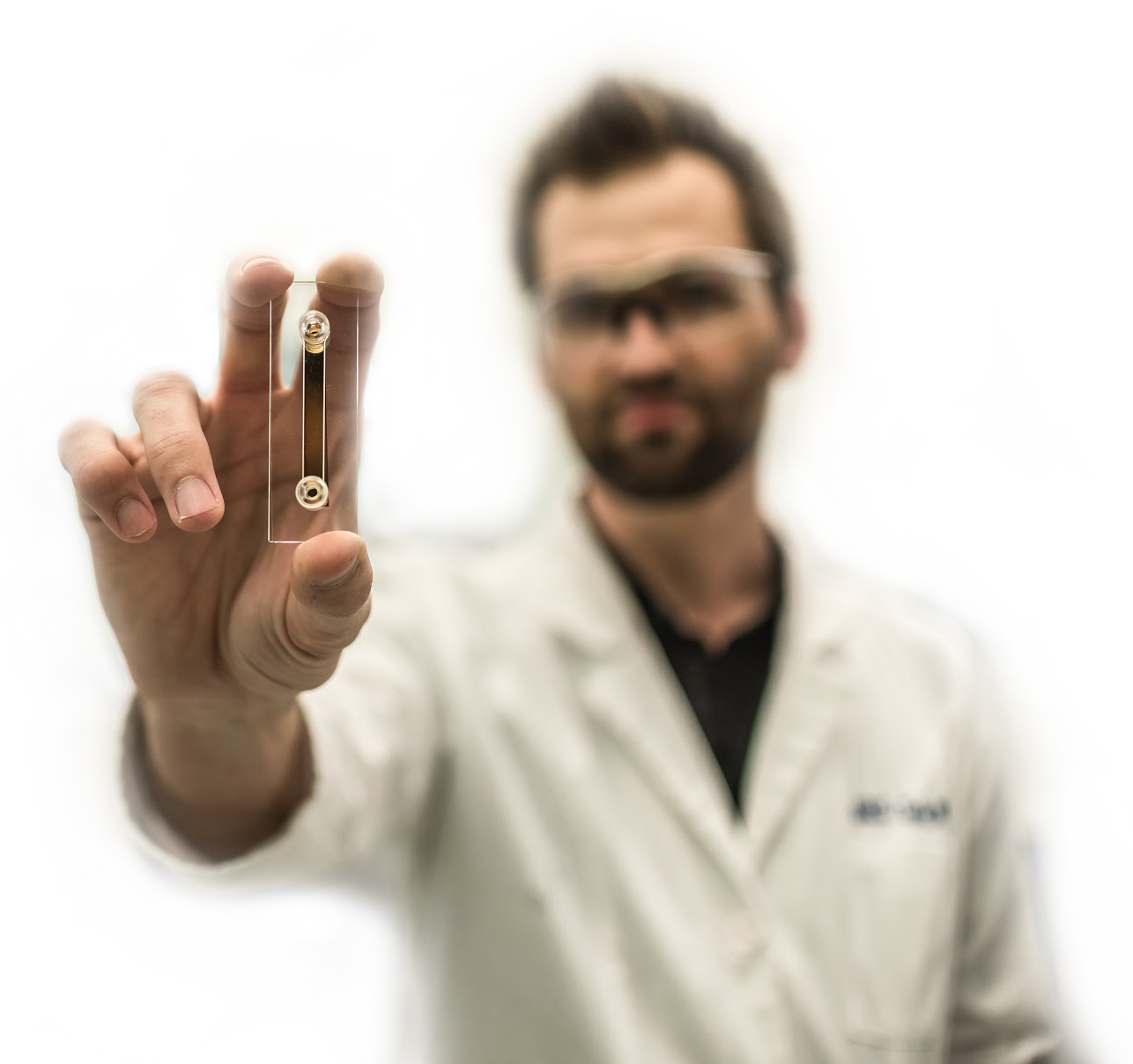
The devices are fabricated at the Joint School of Nanoscience & Nanoengineering (JSNN) in the cleanroom facilities. Layers of metals only a few hundred nanometers thick are deposited onto a glass slide. Then, nanostructured designs 100,000 times thinner than a human hair are carved into the metal surface. A second piece of glass encloses the device, and a syringe is attached to flow samples over the surface through a channel etched in the glass. This flow channel is only a few thousand nanometers in size.
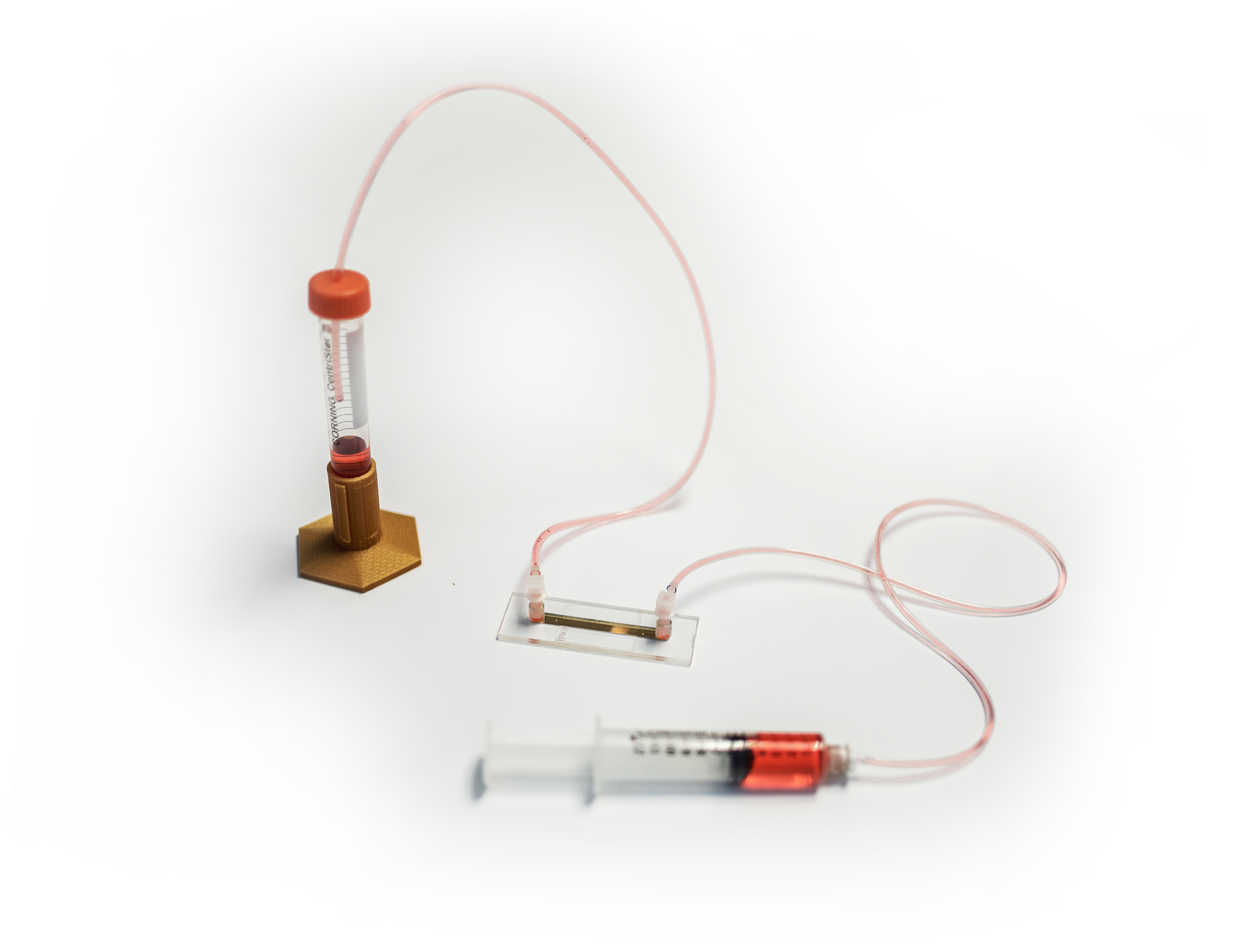
The devices can accommodate any biological sample, such as urine or blood. The sensor works optically, using light to “sense” for biomarkers that bind to the nanostructured surface. But how does the sensor distinguish from one biological molecule to the next? This is done by a form of chemistry called self-assembly, where molecules that act like baseball gloves assemble on the metallic surface. These glove-like molecules, called ligands, are attached to the surface and are designed to only recognize the biomolecules of interest – the “baseballs.” Think about a cop on a busy highway that only pulls over the red convertibles and lets the other cars pass by. This is similar to how our surface-bound ligands work. They “see” all the biological molecules flowing over the surface, but the only ones that stick are the molecules that fit the “glove”.
The ligand in our latest device recognizes retinol-binding protein. Deficiency or excess in retinol-binding protein during pregnancy can cause embryo mortality or developmental deformities, and monitoring its levels is critical. Our work is a first step towards developing a portable retinol-binding protein biosensor – a device could help monitor pregnancies in developing countries, in places where laboratories are essentially nonexistent.
Large benchtop instruments that are commonly used in chemical analysis have million dollar price tags, a lot of moving parts, and are difficult to miniaturize. Prisms, moveable mirrors, temperature control, lasers, fancy optics, and high voltage power supplies make shrinking an analytical instrument very difficult. Using nanoscience is allowing us to overcome these hurdles. For example, the nano-sized surface structures in our devices are smaller than the wavelength of visible light and can essentially break up white light into different wavelengths. As the ligands on our nanostructured surfaces bind target biomarkers, mass is added to the surface of the device, resulting in a change in light. This allows us to detect biomarker presence without the lasers and bulky optics that larger instrumentation requires.
Biosensors have benefits in addition to their portability. By examining samples on-site, they eliminate extra costs and time related to shipping samples to a centralized laboratory. They reduce costs, solvents, and generated waste. With today’s technology, it’s astonishing that blood, water, and other samples are being shipped around the globe. Why not bring the lab to the patient’s bedside?
Basically, we are trying to do for disease diagnostics what the portable glucose meter did for diabetics. Just as computers have gone from the tabletop to the palm of the hand, shrinking chemical instrumentation can also revolutionize the world. These small biological sensors are the future of disease diagnostics and personalized medicine.
By Taylor Mabe, PhD candidate, UNCG Department of Nanoscience, Joint School of Nanoscience and Nanoengineering
Photography by Venky Kollu
Through the Research Perspectives blog, students, staff, and faculty share their successes in research, creative activity, and community and economic engagement with each other and the world at large. Interested in contributing? Click here.